Bone density loss accelerates dramatically in the years around perimenopause. The interventions that protect it must begin before significant loss has occurred. Here is the evidence on the window, the risks, and what actually works.
What Perimenopause Does to Your Bones (And the Window You Cannot Afford to Miss)
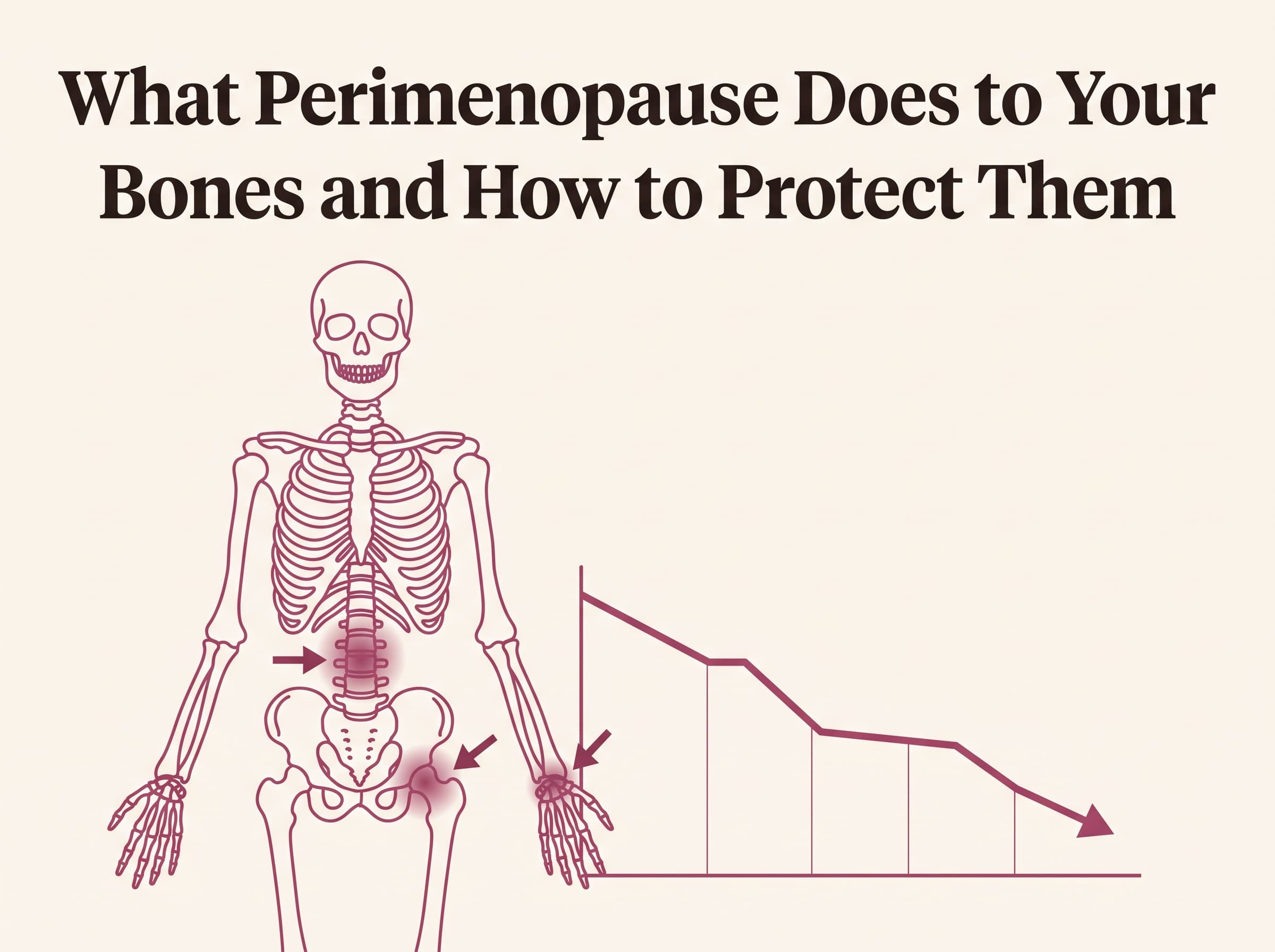
Women lose up to 20 percent of their bone density in the five to seven
years around menopause. This is one of the most significant and most
consistently underappreciated health consequences of the perimenopausal
transition.
It is underappreciated for a straightforward reason: bone loss is invisible.
There is no symptom of losing bone density as it happens. No pain, no
functional impairment, nothing that signals to the woman experiencing it
that the structural integrity of her skeleton is changing in ways that will
have consequences decades later.
By the time osteoporosis is diagnosed — typically after a fracture or a
routine DEXA scan — the loss has already occurred. The window during which
intervention produces the greatest protective effect is the perimenopausal
transition itself, before significant loss has accumulated.
Understanding what is happening, why it is happening, and what has the
strongest evidence for protection is not a concern for women in their 60s.
It is an urgent priority for women in their 40s.
Why Perimenopause Accelerates Bone Loss
The Estrogen-Bone Connection
Estrogen plays a central regulatory role in bone metabolism through its
effects on two cell types: osteoblasts, which build bone, and
osteoclasts, which break bone down.
In the presence of adequate estrogen, osteoblast activity is promoted and
osteoclast activity is suppressed. The net result is maintained or slowly
increasing bone density through the reproductive years.
When estrogen declines and fluctuates in perimenopause, this regulatory
balance shifts. Osteoclast activity increases and osteoblast activity
decreases. Bone resorption begins exceeding bone formation, producing
net bone loss that accelerates as estrogen continues to decline toward
menopause.
The rate of loss is not gradual. The period of most rapid bone loss occurs
in the two years before and three years after the final menstrual period,
with loss rates of 2 to 3 percent per year during this window — compared
to the 0.5 to 1 percent annual loss rate of normal aging before perimenopause.
The Calcium-Vitamin D Interaction
Estrogen also affects calcium metabolism. It supports calcium absorption
from the gut, reduces calcium loss through the kidneys, and promotes the
activity of vitamin D in the bone-building pathway.
As estrogen declines, calcium absorption decreases and urinary calcium loss
increases — meaning the same dietary calcium intake produces less bone
protective effect than it did before perimenopause. This is why the calcium
requirements of perimenopausal women are higher than those of younger women,
and why calcium adequacy without vitamin D adequacy is insufficient.
The Risk Factors That Compound Perimenopausal Bone Loss
Not all perimenopausal women lose bone at the same rate. Several factors
significantly amplify the baseline risk:
- Low body weight — adipose tissue produces estrogen through aromatase
activity, providing some protection; very lean women have less of this buffer - Smoking — directly impairs osteoblast function and reduces estrogen levels
- Excessive alcohol — interferes with calcium absorption and osteoblast activity
- Corticosteroid use — significantly accelerates bone resorption
- Previous fracture after age 40 — the strongest single predictor of
future fracture risk - Family history of osteoporosis or hip fracture — reflects inherited
peak bone mass and bone quality factors - Premature ovarian insufficiency — earlier estrogen decline means
a longer period of accelerated bone loss before the typical menopause age - Low calcium and vitamin D intake — compounds the absorption deficit
produced by estrogen decline
Women lose up to 20% of bone density in the five to seven years around
menopause. This accelerated loss phase is the highest-leverage window for
protective intervention — and it begins in perimenopause, not after menopause.
What Actually Protects Bone in Perimenopause
Resistance Training — The Most Powerful Non-Pharmacological Intervention
Mechanical loading — the stress placed on bone by muscle contraction
and impact — is the primary driver of bone remodeling throughout the lifespan.
Bones adapt to the mechanical demands placed on them by increasing density
and structural strength in loaded areas.
Resistance training applies the specific type of mechanical loading that
stimulates bone remodeling most effectively. Studies consistently show that
progressive resistance training produces measurable increases in bone
mineral density at the hip and spine — the two sites of highest
osteoporotic fracture risk — in perimenopausal and postmenopausal women.
The minimum effective stimulus for bone adaptation: resistance training
two to three times per week with exercises that load the spine and hips
(squats, deadlifts, lunges, overhead pressing) at a challenging intensity
— defined as weight that produces meaningful effort in the eight to twelve
repetition range, not light resistance that can be performed for 20 repetitions.
Impact activity — jumping, running, dancing — provides additional bone
stimulus through ground reaction forces. High-impact activity is not
appropriate for all women, particularly those with pre-existing joint issues,
but walking alone does not provide sufficient mechanical stimulus for bone
adaptation.
Calcium Adequacy — With Important Qualifications
The evidence on calcium supplementation for bone health is more nuanced
than commonly presented.
Dietary calcium from food sources is consistently associated with better
bone outcomes than supplemental calcium and without the cardiovascular concerns
raised by high-dose calcium supplementation in some studies.
The evidence-based targets for perimenopausal women:
- Dietary calcium from dairy, tinned fish with bones, dark leafy greens,
and fortified foods — targeting 1,200mg daily from food sources where possible - Supplemental calcium only to fill the gap when dietary intake is
consistently below 800mg — and in calcium citrate form (better absorbed
than carbonate, particularly in lower stomach acid environments common after 40) - Never more than 500mg supplemental calcium at one time — the gut
cannot absorb more than this per dose and divided dosing produces better
bone outcomes
Vitamin D — At Levels That Actually Support Bone
Vitamin D deficiency is essentially universal in women above the 35th parallel
and common even in women in sunnier climates who use SPF and avoid direct sun
exposure.
The standard laboratory lower limit of 30 ng/mL is not the same as the
optimal level for bone health. The evidence-based optimal range for bone
protection is 50 to 80 ng/mL — a level that most women without deliberate
supplementation do not reach.
The supplemental dose required to reach and maintain this range varies
significantly by starting level, body weight, and sun exposure. A practical
starting point for most perimenopausal women is 2,000 to 4,000 IU of
vitamin D3 daily, with testing at three to six months to confirm levels
have reached the target range.
Vitamin D3 should be taken with vitamin K2 (MK-7 form, 100 to 200mcg daily)
to direct calcium to bone rather than arterial walls — a clinically important
pairing that is underappreciated in standard supplementation advice.
HRT — The Most Effective Intervention for Bone Protection
HRT with transdermal estradiol is the most effective intervention for
preventing perimenopausal bone loss. Estrogen directly addresses the mechanism
of bone loss — the shift in osteoblast-osteoclast balance produced by estrogen
decline — rather than working around it.
Large population studies consistently show that women who use HRT through
perimenopause and early menopause have significantly higher bone density and
significantly lower fracture rates than age-matched non-users. The bone
protective effect is present during HRT use and reverses after discontinuation
as bone resorption accelerates again — supporting the view that HRT addresses
bone protection most effectively as an ongoing treatment rather than a
short-term intervention.
For women with bone loss already identified on DEXA scan, specialist assessment
may recommend additional pharmacological interventions (bisphosphonates,
denosumab, or other bone-specific agents) alongside or instead of HRT,
depending on the degree of loss and individual clinical factors.
What to Do Right Now
The first step for any perimenopausal woman who has not had a recent DEXA
scan is to establish a baseline.
A DEXA scan provides T-scores and Z-scores that quantify bone density relative
to peak bone mass and age-matched peers. This baseline tells you where you
are starting from and allows you to measure whether interventions are
working over time. The scan is low-radiation, takes approximately 20 minutes,
and is the essential foundation for making informed decisions about bone
protective interventions.
Ask your doctor about DEXA scanning at your next appointment, particularly
if you have any of the risk factors listed above. The appropriate time to
establish this baseline is during perimenopause — before significant loss
has occurred — not after.
For the complete guide to perimenopause bone health including DEXA
interpretation, supplement protocols, and exercise specifics, visit
perimenopauseedit.estorealm.com.
Frequently Asked Questions
At what age should perimenopausal women get a DEXA scan?
General guidelines recommend DEXA at 65 for all women, but women with risk
factors — family history of osteoporosis, low body weight, smoking, previous
fracture, or early perimenopause — should be assessed earlier. Many menopause
specialists recommend establishing a baseline DEXA during perimenopause
for any woman with risk factors, to quantify loss and guide protective
interventions before significant damage has occurred.
Does walking protect bone density?
Walking provides some bone stimulus through ground reaction forces but is
not sufficient as a sole bone-protective exercise. The mechanical loading
required for meaningful bone adaptation requires resistance that challenges
the muscular-skeletal system — impact forces from walking are generally below
the threshold that drives significant new bone formation. Walking is valuable
for overall health and fall prevention but should be combined with resistance
training for bone protection.
Can bone density be rebuilt once lost?
Bone density can be increased from a depleted baseline through resistance
training, nutritional optimization, and where appropriate, pharmacological
intervention. However the structural microarchitecture of bone — the
trabecular scaffold that determines fracture resistance independent of
density — is harder to restore once significantly disrupted. This is one
of the primary arguments for early intervention during perimenopause rather
than waiting for significant loss to occur.
Does calcium supplementation cause heart attacks?
Some studies raised concerns about high-dose supplemental calcium and
cardiovascular risk. The concern appears most relevant to high-dose calcium
carbonate supplementation (1,200mg or more daily as a supplement) and is
significantly reduced when calcium comes from food sources, when supplemental
doses are kept below 500mg per dose, and when vitamin K2 is taken alongside
to direct calcium to bone rather than arterial walls. The relevant precaution
is not to avoid calcium but to prioritize food sources and pair supplementation
with K2.
Is joint pain the same as bone loss?
No. Joint pain in perimenopause is primarily an inflammatory process driven
by estrogen withdrawal from joint tissue — estrogen receptors are present
in cartilage and synovial tissue. Bone loss is a structural change in bone
density driven by the osteoblast-osteoclast imbalance described above.
Both are driven by declining estrogen and both are addressed by HRT, but
they are distinct processes with distinct mechanisms and distinct sites.
This article is for educational purposes only and does not constitute medical
advice. Please consult a qualified healthcare provider for bone health
assessment and treatment guidance specific to your health history.
More from The Perimenopause Edit
The Nutrition Formula That Actually Works for Women After 40 in Perimenopause
Nutritional requirements change in perimenopause in specific, documented ways. The formula that works after 40 is not a clean eating plan. It is a targeted approach addressing the hormonal, metabolic, and structural changes that make standard dietary advice insufficient.
The Neurochemical Reason Your Mood and Anxiety Changed in Perimenopause
New-onset anxiety in your 40s with no clear life trigger is not a psychological problem. It is a neurochemical one. Here is the mechanism and what addresses it at the level it is actually occurring.
Why Your Weight Changed After 40 Even Though Nothing Else Did
Eating the same. Moving the same. Weight redistributing anyway. The explanation is hormonal and the interventions that work address the hormonal environment, not just the calories.
The next the perimenopause edit essay in your inbox.
One careful letter, every Sunday. Free.